6.5 Nerves, hormones, and homeostasis
6.5.1 Central nervous system
|
The nervous system is a
network of specialized cells that communicate information about an
organism's surroundings and itself. It processes this information and
causes reactions in other parts of the body. It is composed of
neurons
and other specialized cells called
glia, that aid in the function of the neurons. The nervous system is
divided broadly into two categories: the
peripheral nervous system and the
central nervous system. Neurons generate and conduct
impulses
between and within the two systems. |
 |
1. Central Nervous System (CNS)
a. Structures of
the CNS:
-Brain
-Spinal cord
b. Function:
The CNS coordinates and
interprets information to determine the best response
2.
Peripheral Nervous System (PNS)
a. Consist of Cranial nerves
(12 pairs) &
Spinal nerves: (31 pairs)
- Carry nerve impulses to and from
the spinal cord to body parts not served by the cranial nerves. Sensory
(afferent) nerves bring sensory inputs into the CNS and motor (efferent)
nerves takes signals back out.
b. Can be separated into:
1.
Somatic nervous system -
Carries nerve impulses to the skeletal muscles, joints and skin
2. Autonomic nervous system (ANS)
-Carries nerve impulses to the
smooth muscles of internal organs and to glands without conscious
thought
Two subdivisions of autonomic nervous system
:
-Sympathetic system
- Controls
"fight or flight" responses. Its neurotransmitter is norepinephrine
(or noradrenaline). "Inducible system"
-Parasympathetic
system -Controls
those responses associated with a relaxed state. Its neurotransmitter is acetylcholine (Ach).
|
| Nervous system animation:
http://health.howstuffworks.com/adam-200011.htm
|
|
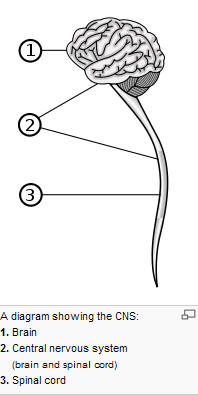
| Central Nervous
System |
|
|
The central nervous system (CNS) is the part of the
nervous system that functions to coordinate the activity of all
parts of the bodies of multicellular organisms. The CNS contains the
majority of the nervous system is contained within the
dorsal cavity, with the
brain in
the
cranial cavity and the
spinal cord in the
spinal cavity. The brain is protected by the skull, while the spinal
cord is protected by the vertebrae. Together with the
peripheral nervous system it has a fundamental role in the control
of
behavior.
The CNS is covered by the
meninges, a three layered protective coat. consisting of: the
dura mater, the
arachnoid mater, and the
pia
mater. The primary function of the meninges and of the
cerebrospinal fluid is to protect the
central nervous system
 |
| Peripheral
Nervous System |
|
 |
The peripheral nervous system (PNS) resides or extends
outside the
central nervous system (CNS). The main function of the PNS is to
connect the CNS to the limbs and organs. Unlike the central nervous
system, the PNS is not protected by
bone or by
the
blood-brain barrier, leaving it exposed to
toxins
and mechanical injuries. The peripheral nervous system is divided into
the
somatic nervous system and the
autonomic nervous system |
|
Neurons |
|
 |
Neurons are electrically excitable cells in the nervous
system that process and transmit information by
electrochemical
signalling. Neurons are the core components of the
brain, the vertebrate spinal cord, the invertebrate
ventral nerve cord, and the peripheral nerves. They use
electrochemical signals and
neurotransmitters to transmit impulses from one
neuron to the next. A number of different types of
neurons exist:
1. Sensory
neurons
(Afferent Neurons) respond to
touch, sound, light and numerous other stimuli effecting
sensory organs and send signals to the CNS, away from
the peripheral system. Unlike neurons of the central
nervous system, whose inputs come from other neurons,
sensory neurons are activated by physical modalities
such as light, sound, temperature, chemical stimulation,
etc.
2. motor
neurons (Efferent Neurons)
receive signals from the CNS and carry this to the
peripheral system, causing muscle contractions and
affecting glands.
3.
Interneurons are multipolar neurons that connect
neurons to other neurons within the brain and spinal
cord. The reside entirely within the CNS
|
|
|
|
6.5.2 Motor neuron
In
vertebrates, the term
motor neuron (or
motoneuron)
classically applies to
neurons located in the
central nervous system (or CNS) that project their
axons outside the CNS and directly or indirectly control
muscles. Motor neuron is often associated with
efferent neuron, primary neuron, or
alpha motor neurons.
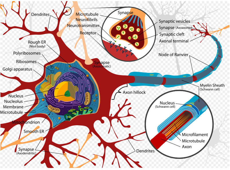 |
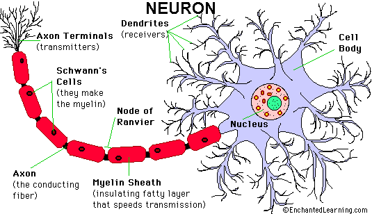
- The
soma is the central part of the
neuron. It contains the
nucleus of the cell, and
therefore is where most
protein synthesis occurs. The
nucleus ranges from 3 to 18
micrometers in diameter.[11]
- The
dendrites of a neuron are
cellular extensions with many
branches, and metaphorically this
overall shape and structure is
referred to as a dendritic tree.
This is where the majority of input
to the neuron occurs. Information
outflow (i.e. from dendrites to
other neurons) can also occur, but
not across chemical synapses; there,
the back flow of a nerve impulse is
inhibited by the fact that an axon
does not possess chemoreceptors and
dendrites cannot secrete
neurotransmitter chemicals. This
unidirectionality of a chemical
synapse explains why nerve impulses
are conducted only in one direction.
- The
axon is a finer, cable-like
projection which can extend tens,
hundreds, or even tens of thousands
of times the diameter of the soma in
length. The axon carries nerve
signals away from the soma (and also
carries some types of information
back to it). Many neurons have only
one axon, but this axon may - and
usually will - undergo extensive
branching, enabling communication
with many target cells. The part of
the axon where it emerges from the
soma is called the
axon hillock. Besides being an
anatomical structure, the axon
hillock is also the part of the
neuron that has the greatest density
of voltage-dependent sodium
channels. This makes it the most
easily-excited part of the neuron
and the spike initiation zone for
the axon: in neurological terms it
has the most negative
action potential threshold.
While the axon and axon hillock are
generally involved in information
outflow, this region can also
receive input from other neurons.
|
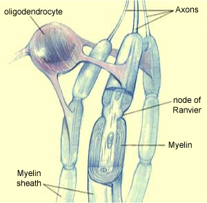 |
Axons often have an insulating sheath
that lets nerve impulses travel faster. This sheath is made of a fatty
substance called myelin, which consists of glial cell membranes wrapped
around the axon. The myelin of the neurons in the brain is composed of
oligodendrocytes, while that of the neurons in the peripheral nervous
system is composed of Schwann cells. The myelin sheath does not
cover the entire axon; it leaves small sections uncovered. These small
exposed sections are called nodes of Ranvier. They are spaced from 0.2
to 2 millimeters apart.
The reason that the myelin sheath speeds up neural conduction is that
the action potentials literally jump from one node of Ranvier to the
next. In fact, these nodes are the only places where the ion exchanges
that generate the action potential can take place.
This process is called saltatory conduction (from the Latin saltare,
meaning “to jump”), as opposed to the much slower, continuous
propagation that occurs in non-myelinated axons.
|
6.5.3 Nerve impulses
nerve impulse animation:
http://highered.mcgraw-hill.com/sites/0072495855/student_view0/chapter14/animation__the_nerve_impulse.html
6.5.4 Resting and action potentials
An action potential is a self-regenerating
wave of
electrochemical activity that allows
nerve cells to carry a signal over a distance. It is the primary electrical
signal generated by nerve cells, and arises from changes in the
permeability of the nerve cell's axonal
membranes to specific
ions. Action potentials (also known as nerve impulses or spikes)
are pulse-like waves of
voltage that
travel along several types of
cell
membranes.[1]
The best-understood example of an action potential is generated on the membrane
of the axon of a
neuron, but
also appears in other types of excitable
cells, such as
cardiac muscle cells, and even
plant cells.
Neuron potentials animations:
http://bcs.whfreeman.com/thelifewire/content/chp44/4401s.swf
Studying action potentials animation:
http://www.sumanasinc.com/webcontent/animations/content/action_potential.html
Channel gating during action potential animation:
http://www.blackwellpublishing.com/matthews/channel.html
Sodium-Potassium Exchange:
http://highered.mcgraw-hill.com/sites/0072437316/student_view0/chapter45/animations.html#
6.5.5 Nerve impulse
Nerve impulse (myelinated vs. nonmyelinated neurons) animation:
http://www.blackwellpublishing.com/matthews/actionp.html
Action Potential Propagation in an Unmyelinated Axon:
http://highered.mcgraw-hill.com/sites/0072437316/student_view0/chapter45/animations.html#
6.5.6 Synaptic transmission
Synaptic transmission animation:
http://highered.mcgraw-hill.com/sites/0072495855/student_view0/chapter14/animation__transmission_across_a_synapse.html
neuromuscular synapse animation:
http://www.sumanasinc.com/webcontent/animations/content/synaptictransmission.html
synaptic vesicle release animation:
http://www.blackwellpublishing.com/matthews/nmj.html
Comparison of direct and indirect neurotransmitter actions:
http://www.blackwellpublishing.com/matthews/neurotrans.html
6.5.7 Endocrine system
The endocrine system is a system of small organs that involve the
release of
extracellular signaling molecules known as
hormones. The
endocrine system is instrumental in regulating
metabolism,
growth, development and puberty, and
tissue function and also plays a part in determining
mood.[1]
The field of study that deals with disorders of endocrine glands is
endocrinology, a branch of the wider field of internal medicine.
Hormones:
http://users.rcn.com/jkimball.ma.ultranet/BiologyPages/H/Hormones.html
Hormonal communication animation:
http://highered.mcgraw-hill.com/sites/0072495855/student_view0/chapter20/animation__hormonal_communication.html
Endocrine pathology (images):
http://library.med.utah.edu/WebPath/ENDOHTML/ENDOIDX.html
Hormone action animations:
http://www.wisc-online.com/objects/index_tj.asp?objID=AP13704
6.5.8 Homeostasis
Homeostasis animation:
http://health.howstuffworks.com/adam-200092.htm
Site:
http://www.biology-online.org/4/1_physiological_homeostasis.htm
6.5.9 Monitoring
Positive & negative feedback animation:
http://highered.mcgraw-hill.com/sites/0072495855/student_view0/chapter28/animation__positive_and_negative_feedback__quiz_2_.html
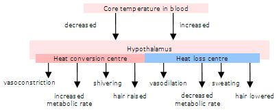
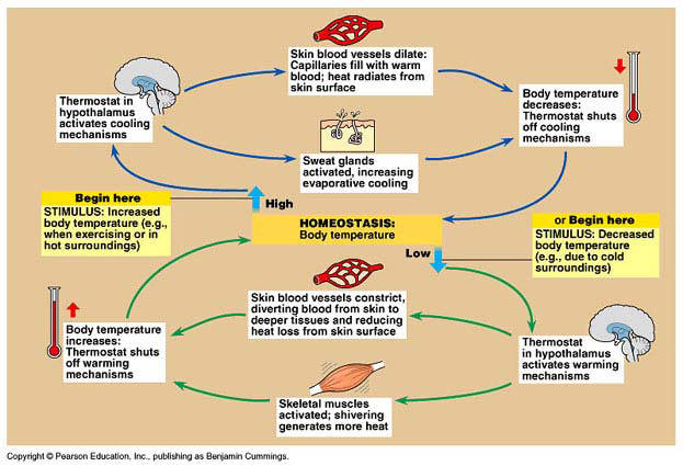
6.5.10 Body temperature & homeostasis
One of the most important examples of
homeostasis is the regulation of body temperature. Not all animals can do this
physiologically. Animals that maintain a fairly constant body temperature (birds
and mammals) are called
endotherms,
while those that have a variable body temperature (all others) are called
ectotherms.
Endotherms normally maintain their body temperatures at around 35 - 40°C, so are
sometimes called
warm-blooded
animals, but in fact ectothermic animals can also have
very warm blood during the day by basking in the sun, or by extended muscle
activity 9e.g. bumble bees, tuna). The difference between the two groups is thus
that endothermic animals use
internal
corrective mechanisms, whilst ectotherms use
behavioural
mechanisms (e.g. lying in the sun when cold, moving
into shade when hot). Such mechanisms can be
very
effective, particularly when coupled with
internal
mechanisms to ensure that the temperature of the blood
going to vital organs (brain, heart) is kept constant. We use
both!
In humans, body temperature is controlled by the
thermoregulatory centre in the
hypothalamus.
It receives input from
two sets
of
thermoreceptors:
receptors in
the hypothalamus itself monitor the temperature of the
blood as it passes through the brain (the
core
temperature), and
receptors in
the skin (especially on the trunk) monitor the
external
temperature.
Both
sets of information are needed so that the body can make
appropriate adjustments. The thermoregulatory centre sends impulses to several
different effectors to adjust body temperature:
6.5.11 Glucose & homeostasis
Glucose levels animation:
http://highered.mcgraw-hill.com/sites/0072495855/student_view0/chapter20/animation__blood_sugar_regulation_in_diabetics.html
Glucose levels tutorial:
http://www.wisc-online.com/objects/index_tj.asp?objID=AP15004
6.5.12 Diabetes
 |
Diabetes mellitus is a
syndrome of disordered
metabolism, usually due to a combination of
hereditary and environmental causes, resulting in abnormally high
blood sugar levels (hyperglycemia).
Blood glucose levels are controlled by a complex interaction of multiple
chemicals and hormones in the body, including the
hormone
insulin
made in the
beta
cells of the
pancreas. Diabetes mellitus refers to the group of diseases that
lead to high blood glucose levels due to defects in either insulin
secretion or insulin action in the body. Diabetes develops due to a
diminished production of
insulin
(in
type 1) or resistance to its effects (in
type 2 and
gestational). Both lead to hyperglycemia, which largely causes
the acute signs of diabetes:
excessive urine production, resulting compensatory
thirst and increased fluid intake, blurred vision, unexplained
weight loss,
lethargy, and changes in energy
metabolism.
All forms of diabetes have been treatable since
insulin
became medically available in 1921, but there is no cure. The
injections by a
syringe,
insulin pump, or
insulin pen deliver insulin, which is a basic
treatment of type 1 diabetes.
Type 2 is managed with a combination of
dietary treatment,
exercise,
medications and insulin supplementation.
Diabetes and its treatments can cause many
complications.
Acute complications ( hypoglycemia,
ketoacidosis, or
nonketotic hyperosmolar coma) may occur if the
disease is not adequately controlled. Serious long-term
complications include
cardiovascular disease (doubled risk),
chronic renal failure,
retinal damage (which can lead to
blindness),
nerve damage (of several kinds), and microvascular
damage, which may cause
erectile dysfunction and poor wound healing. Poor
healing of wounds, particularly of the feet, can lead to
gangrene, and possibly to
amputation. Adequate treatment of diabetes, as well
as increased emphasis on
blood pressure control and lifestyle factors (such
as not
smoking and maintaining a healthy
body weight), may improve the risk profile of most
of the chronic complications. In the developed world,
diabetes is the most significant cause of adult
blindness in the non-elderly and the leading cause of
non-traumatic amputation in adults, and
diabetic nephropathy is the main illness requiring
renal dialysis in the United States
|
Type 1 diabetes mellitus is characterized by loss of the
insulin-producing
beta cells of the
islets of Langerhans in the pancreas leading to a deficiency
of insulin. This type of diabetes can be further classified as
immune-mediated or idiopathic. The majority of type 1 diabetes
is of the immune-mediated variety, where beta cell loss is a
T-cell mediated
autoimmune attack.
[3]
There is no known preventive measure which can be taken against
type 1 diabetes; it is about 10% of diabetes mellitus cases in
North America and Europe (though this varies by geographical
location), and is a higher percentage in some other areas. Most
affected people are otherwise healthy and of a healthy weight
when onset occurs. Sensitivity and responsiveness to insulin are
usually normal, especially in the early stages. Type 1 diabetes
can affect children or adults but was traditionally termed
"juvenile diabetes" because it represents a majority of the
diabetes cases in children. The term "type 1 diabetes" has
universally replaced several former terms, including
childhood-onset diabetes, juvenile diabetes, and
insulin-dependent diabetes mellitus (IDDM).
Type 2 diabetes mellitus is characterized differently and is due to
insulin resistance or reduced insulin sensitivity, combined with
relatively reduced insulin secretion which in some cases becomes
absolute. The defective responsiveness of body tissues to
insulin almost certainly involves the
insulin receptor in cell membranes. However, the specific
defects are not known. Diabetes mellitus due to a known specific
defect are classified separately. Type 2 diabetes is the most
common type. The term "type 2 diabetes" has replaced several
former terms, including adult-onset diabetes, obesity-related
diabetes, and non-insulin-dependent diabetes mellitus (NIDDM).
Insulin interaction:
http://www.abpischools.org.uk/page/modules/hormones/horm6.cfm
Type II diabetes animation:
http://www.healthscout.com/animation/1/34/main.html
Agrosy animations:
http://www.argosymedical.com/medical_ani_sys/nervous.html