
6.2 Transport system
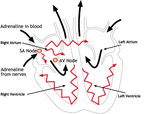
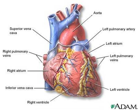
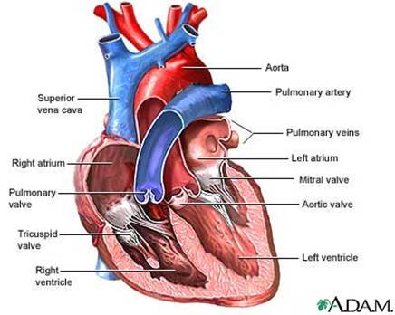
6.2.1- heart: chambers, blood vessels, valves, rout of blood
|
|
||
|
|
||
|
||
|
|
||
6.2.2 Coronary arteries
 |
|
|
6.2.3
Systole
Diastole
6.2.4 Heart beat
 |
|
|
|
The sinoatrial node (abbreviated SA node or SAN, also called the sinus node) is the impulse generating (pacemaker) tissue located in the right atrium of the heart, and thus the generator of sinus rhythm. It is a group of cells positioned on the wall of the right atrium, near the entrance of the superior vena cava. These cells are modified cardiac myocytes. Though they possess some contractile filaments, they do not contract. Although all of the heart's cells possess the ability to generate the electrical impulses (or action potentials) that trigger cardiac contraction, the sinoatrial node is what normally initiates it, simply because it generates impulses slightly faster than the other areas with pacemaker potential. Because cardiac myocytes, like all muscle cells, have refractory periods following contraction during which additional contractions cannot be triggered, their pacemaker potential is overridden by the sinoatrial node. In the absence of extrinsic neural and hormonal control, cells in the SA node, situated in the upper right corner of the heart, will naturally discharge (create action potentials) at about 70-80 beats/minute.[2] Because the sinoatrial node is responsible for the rest of the heart's electrical activity, it is sometimes called the primary pacemaker. If the SA node does not function, or the impulse generated in the SA node is blocked before it travels down the electrical conduction system, a group of cells further down the heart will become the heart's pacemaker. These cells form the atrioventricular node (AV node), which is an area between the atria and ventricles, within the atrial septum. The atrioventricular node (abbreviated AV node) is a part of electrical control system of the heart that co-ordinates heart rate. It electrically connects atrial and ventricular chambers. [1] The AV node is an area of specialized tissue between the atria and the ventricles of the heart, specifically in the posteroinferior region of the interatrial septum near the opening of the coronary sinus, which conducts the normal electrical impulse from the atria to the ventricles. The AV node is quite compact (~1 x 3 x 5 mm).[2] It is located at the center of Koch's Triangle--a triangle enclosed by the septal leaflet of the tricuspid valve, the coronary sinus, and the membraneous part of the interatrial septum.[3]
|
||
 |
||
| Cardiac cycle is the term referring to all or any of the events related to the flow or pressure of blood that occurs from the beginning of one heartbeat to the beginning of the next.[1] The frequency of the cardiac cycle is the heart rate. Every single 'beat' of the heart involves three major stages: atrial systole, ventricular systole and complete cardiac diastole. The term diastole is synonymous with relaxation of a muscle. Throughout the cardiac cycle, the blood pressure increases and decreases. The cardiac cycle is coordinated by a series of electrical impulses that are produced by specialized heart cells found within the sino-atrial node and the atrioventricular node. The cardiac muscle is composed of myocytes which initiate their own contraction without help of external nerves (with the exception of modifying the heart rate due to metabolic demand). | ||
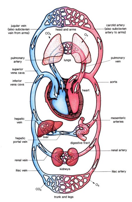
6.2.5 Arteries, capillaries and veins
 |
||
 |
||
 |
||
 |
||
6.2.6 Blood composition
 |
In vertebrates, it is composed of blood cells suspended in a liquid called blood plasma. Plasma, which comprises 55% of blood fluid, is mostly water (90% by volume [1] ), and contains dissolved proteins, glucose, mineral ions, hormones, carbon dioxide (plasma being the main medium for excretory product transportation), platelets and blood cells themselves. The blood cells present in blood are mainly red blood cells (also called RBCs or erythrocytes) and white blood cells, including leukocytes and platelets (also called thrombocytes). The most abundant cells in vertebrate blood are red blood cells. These contain hemoglobin, an iron-containing protein, which facilitates transportation of oxygen by reversibly binding to this respiratory gas and greatly increasing its solubility in blood. In contrast, carbon dioxide is almost entirely transported extracellularly dissolved in plasma as bicarbonate ion
|
|||||||||||||||||||||||||||||||||||||||||||||||||||||||||||||||||||||||||||||||||||||||||||||||||||||||||||
 |
Red blood cells are the most common type of
blood cell and the
vertebrate body's principal means of delivering
oxygen to the body tissues via the
blood. The cells are filled with
hemoglobin, a
biomolecule that can bind to oxygen. They take up
oxygen in the
lungs or
gills and release it while squeezing through the
body's
capillaries. The blood's red color is due to the
color of hemoglobin. In humans, red blood cells develop
in the
bone marrow, take the form of flexible biconcave
disks, lack a
cell nucleus, subcellular
organelles and the ability to
synthesize protein, and live for about 120 days.[1]
Red blood cells are also known as RBCs, red blood corpuscles (an archaic term), haematids or erythrocytes (from Greek erythros for "red" and kytos for "hollow", with cyte translated as "cell" in modern usage). The capitalized term Red Blood Cells is the proper name in the US for erythrocytes in storage solution used in transfusion medicine.[2]
|
|||||||||||||||||||||||||||||||||||||||||||||||||||||||||||||||||||||||||||||||||||||||||||||||||||||||||||
|
||||||||||||||||||||||||||||||||||||||||||||||||||||||||||||||||||||||||||||||||||||||||||||||||||||||||||||
| White blood cells (WBCs),
or leukocytes (also spelled "leucocytes"), are
cells of the
immune system defending the body against both
infectious disease and foreign materials. Five[1]
different and diverse types of leukocytes exist, but they are all
produced and derived from a
multipotent cell in the
bone marrow known as a
hematopoietic stem cell. Leukocytes are found throughout the body,
including the
blood and
lymphatic system.[2] The number of leukocytes in the blood is often an indicator of disease. There are normally between 4×10⁹ and 11×109 white blood cells in a litre of blood, making up approximately 1% of blood in a healthy adult.[3] In conditions such as leukemia, the number of leukocytes is higher than normal, and in leukopenia, this number is much lower. The physical properties of leukocytes, such as volume, conductivity, and granularity, may change due to activation, the presence of immature cells, or the presence of malignant leukocytes in leukemia.
|
||||||||||||||||||||||||||||||||||||||||||||||||||||||||||||||||||||||||||||||||||||||||||||||||||||||||||||
 |
Platelets, or thrombocytes, are small, irregularly shaped anuclear cells, 2-4µm in diameter, which are derived from fragmentation of precursor megakaryocytes. The average life span of a platelet is between 8 and 12 days. Platelets play a fundamental role in hemostasis and are a natural source of growth factors[1]. They circulate in the blood of mammals and are involved in hemostasis leading to the formation of blood clots. Like red blood cells, platelets have no nucleus | |||||||||||||||||||||||||||||||||||||||||||||||||||||||||||||||||||||||||||||||||||||||||||||||||||||||||||
6.2.7 Solutes in blood
 |
Antibodies (also known as immunoglobulins[1], abbreviated Ig) are gamma globulin proteins that are found in blood or other bodily fluids of vertebrates, and are used by the immune system to identify and neutralize foreign objects, such as bacteria and viruses. They are typically made of basic structural units—each with two large heavy chains and two small light chains—to form, for example, monomers with one unit, dimers with two units or pentamers with five units. Antibodies are produced by a kind of white blood cell called a plasma cell. There are several different types of antibody heavy chains, and several different kinds of antibodies, which are grouped into different isotypes based on which heavy chain they possess. Five different antibody isotypes are known in mammals, which perform different roles, and help direct the appropriate immune response for each different type of foreign object they encounter.[2 | |
 |
||
|
Three kinds of chemical signaling can be distinguished;
http://users.rcn.com/jkimball.ma.ultranet/BiologyPages/H/Hormones.html
|
Hormones (from
Greek ὁρμή - "impetus") are
chemicals released by
cells that affect cells in other parts of the body.
Only a small amount of hormone is required to alter cell
metabolism. It is essentially a chemical messenger
that transports a signal from one cell to another. All
multicellular organisms produce hormones;
plant hormones are also called
phytohormones. Hormones in
animals are often transported in the blood. Cells
respond to a hormone when they
express a specific
receptor for that hormone. The hormone binds to the
receptor
protein, resulting in the activation of a
signal transduction mechanism that ultimately leads
to cell type-specific responses.
Endocrine hormone molecules are secreted (released) directly into the bloodstream, while exocrine hormones (or ectohormones) are secreted directly into a duct, and from the duct they either flow into the bloodstream or they flow from cell to cell by diffusion in a process known as paracrine signalling
|
|
Chemical classes of hormonesVertebrate hormones fall into three chemical classes:
|
||


